


GYNAECOLOGIC ULTRASOUND SCANS

Ultrasounds are high-frequency sound waves transmitted by the transducer probe of an ultrasound machine and as they reflect off the body tissue interface, they display an image of the area their beam has focused.
Moreover, blood flow and direction in the scanned tissues is monitored with a color Doppler scanner.
It is particularly important to know that ultrasound is harmless both to you and your baby and may be repeated without any risk as frequently as advised by your doctor.
In the Gynecologic Ultrasound Scan Department you can perform a full examination of lesser pelvis area with an abdominal and transvaginal ultrasound scan and check blood flow of the uterus and relevant parts with a Doppler scan.
In particular, the following can be examined:
- Uterus shape, size, flexion and version and any uterine abnormalities (arcuate, bicornuate, didelphys uterus).
- Myometrial composition and detection of any existing uterus fibroids.
- Endometrium, its thickness and composition and detection of any endometrial polyps.
- Ovaries for cysts or other physiological or pathological conditions, such as endometriosis, ovarian masses, abscesses.
- Determine the number and size of ovarian follicles for ovulation control.
- Assessment of ovarian vascularity with Doppler scan, especially in cases of investigation for masses and in cases of a possible cyst torsion.
- Fallopian tubes, in cases of possible inflammation (hydrosalpinx, pyosalpinx).
- Soft particles and surrounding areas for edemas or fluid that may mean infections or other pathological conditions.
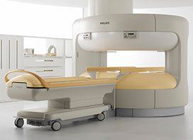
State-of-the-art ultrasound equipment is used for all above tests, with 3D vaginal and abdominal probes for stereoscopic imaging.
A doctor of the department is always available in case of emergency.